Dural arteriovenous fistulas (DAVFs) are abnormal connection between extracranial arteries and intracranial veins, leading to pathological shunting of arterial blood flow to the cerebral veins that are consequently exposed to a too high flow and pressure. When this arterial flow refluxes to veins on the surface of the brain, the affected veins are at risk of rupture. DAVFs with a risk of rupture need to be treated before rupture occurs, with either endovascular embolization, microsurgery, or sometimes stereotactic radiosurgery. DAVFs are not innate but develop during life. Moreover, DAVFs are dynamic lesions that may grow or even spontaneously regress during follow-up. Our aim is to study the pathobiology of DAVFs, to better understand their untreated clinical course, treatment indications, and to possibly develop novel biological therapies that could be used as an option to more invasive procedures, especially when the DAVF does not cause an imminent risk of rupture.
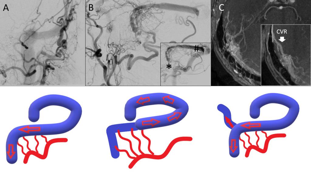
Figure 8. Digital substraction angiography (DSA) examples of DAVFs draining to the transverse and sigmoid sinus with antegrade filling of the sinus (Cognard 1)(A, coronal view. Note filling of the ipsilateral transverse and sigmoid sinus) and with retrograde flow to the transverse sinus (Cognard 2a) (B, sagittal view. Note that the ipsilateral sigmoid sinus (*) is not filling but instead the drainage is to the contralateral sigmoid sinus through retrograde filling of the transverse sinus (#)). Retrograde filling of the sinus with cortical venous reflux, CVR) (Cognard 2a+b) is demonstrated in with MRA in C. Schematic illustrations of the corresponding DAVFs are provided in the lower row. From the book chapter “Stereotactic radiotherapy for dural arteriovenous fistulas” by Frösen J and Lindren AE on the book CyberKnife NeuroRadiosugery: A Practical Guide edited by A.Conti et al. Springer, 2020.
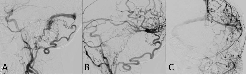
Figure 9. Example of a DAVF that progressed despite radiotherapy. Digital substraction angiography (DSA) prior to radiotherapy (A) showing a simple retrosigmoid DAVF with antegrade draining to the transverse sinus and no cortical venous reflux. A single fraction of radiotherapy with 20Gy margin dose was given. In a control DSA 3 years after the radiotherapy, the DAVF has progressed with now retrograde flow and cortical venous reflux (B: lateral view, C: coronal view). From the book chapter “Stereotactic radiotherapy for dural arteriovenous fistulas” by Frösen J and Lindren AE on the book CyberKnife NeuroRadiosugery: A Practical Guide edited by A.Conti et al. Springer, 2020.