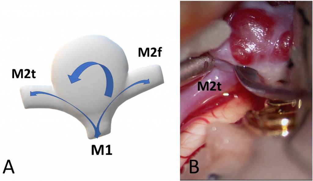
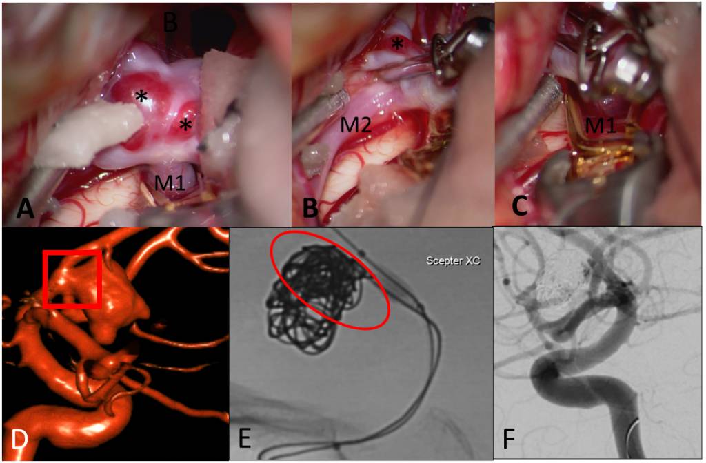
Intracranial aneurysms (IA) are pathological dilatations of large cerebral arteries that run on the surface of the brain in the subarachnoid space. IAs are not innate but develop during life in a relatively large proportion of the population (estimated prevalence >3% in the past middle-aged population). Although only a very small proportion of existing IAs rupture annually (incidence of rupture approx. 0.1/1000 vs. IA prevalence of aneurysms more than 20/1000), the consequences of IA rupture are often devastating with a mortality around 50% and high morbidity among the survivors. Given the high mortality and morbidity related to IA rupture, the best treatment for it is prevention. This, however, would require accurate identification of rupture-prone aneurysms before rupture occurs. The stochastic nature of IA rupture, and the fact that IAs may stay unchanged for years and then suddenly rupture, make this task very demanding.


* Key publications:
Frösen J, Cebral J, Robertson AM, Aoki T. Flow-induced, inflammation-mediated arterial wall remodeling in the formation and progression of intracranial aneurysms. Neurosurg Focus. 2019 Jul 1;47(1):E21. doi: 10.3171/2019.5.FOCUS19234. PMID: 31261126; PMCID: PMC7193287.
Frösen J. Smooth muscle cells and the formation, degeneration, and rupture of saccular intracranial aneurysm wall–a review of current pathophysiological knowledge. Transl Stroke Res. 2014 Jun;5(3):347-56. doi: 10.1007/s12975-014-0340-3. Epub 2014 Apr 1. PMID: 24683005.
Frösen J, Tulamo R, Paetau A, Laaksamo E, Korja M, Laakso A, Niemelä M, Hernesniemi J. Saccular intracranial aneurysm: pathology and mechanisms. Acta Neuropathol. 2012 Jun;123(6):773-86. doi: 10.1007/s00401-011-0939-3. Epub 2012 Jan 17. PMID: 22249619.
Frösen J, Piippo A, Paetau A, Kangasniemi M, Niemelä M, Hernesniemi J, Jääskeläinen J. Remodeling of saccular cerebral artery aneurysm wall is associated with rupture: histological analysis of 24 unruptured and 42 ruptured cases. Stroke. 2004 Oct;35(10):2287-93. doi: 10.1161/01.STR.0000140636.30204.da. Epub 2004 Aug 19. PMID: 15322297.
