
The overarching goal of our research is to increase the present understanding of the visual system in health and in different disease conditions. Our main interest is in the neural retina and in the underlying retinal pigment epithelium (RPE), which keeps the retina alive.
We use electrophysiological and imaging techniques together with cell biology tools to study the functions of these tissues and to determine their interactions. We focus on the functioning of the different ion channels present in the RPE, and our ultimate aim is to dissect out the role of these ion channels in the physiology and pathophysiology of the retina.
With state-of-the-art technology, multidisciplinary research environment and our possibility to use stem cell derived RPE, we can also increase our knowledge on different retinal degenerative diseases and aid the development of novel retinal therapies (e.g. transplantation therapies).
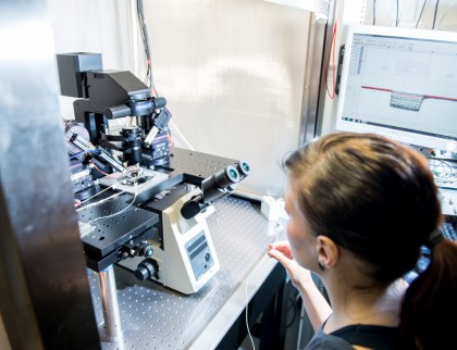 Patch Clamp Technique Patch Clamp TechniqueWith patch clamp technique, we study the functioning of the different ion channels in RPE. Recordings are mainly carried out in whole cell or perforated patch configuration. In our setups we can combine the recordings with simultaneous imaging methods, e.g. using calcium sensitive dyes. |
|
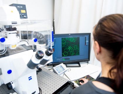 Confocal Microscopy Confocal MicroscopyOur institute is equipped with a multitude of imaging systems including confocal microscopes. We use these systems to investigate the localization of different ion channels and other proteins in retina and RPE both in fixed and live cells. |
|
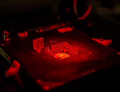 Microelectrode Array (MEA) Microelectrode Array (MEA)We use microelectrode array (MEA) to record the electrical activity of retinal neutrons. Our conventional MEA system allows the recordings of both photoreceptor and ganglion cell activity with moderate resolution (60 electrodes, electrode spacing 200µm and diameter 30µm), whereas our new CMOS-MEA system makes possible the ganglion cell |
|
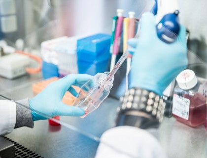 Cell Culturing Cell CulturingWe use cultures of stem cell derived RPE and immortalized cell lines together with freshly isolated tissue in our studies. |
Photos: Miika Fadjukov