Engineered cardiovascular model saved the rat puppies
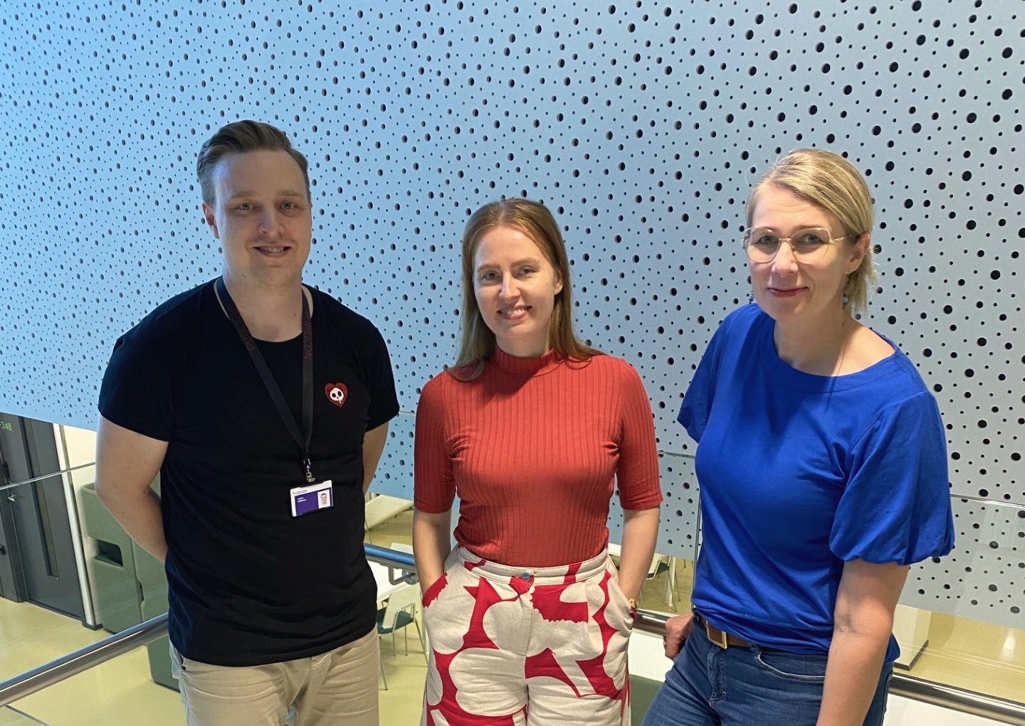
“This work has a story reflecting the progress in science”, says the main author Hanna Vuorenpää.
“In 2014, the first publication on cardiovascular model with neonatal rat cardiomyocytes and a vascular-like network came out. Although rat cardiomyocytes, in other words heart muscle cells were functional in the system, it was evident that they do not reflect human biology. Furthermore, we did not want to sacrifice those small rat puppies anymore”.
In 2017, stem cell technology was already blooming, and the researchers were able to replace neonatal rat cardiomyocytes with human reprogrammed stem cell derived cardiomyocytes, reporting a completely human cell based cardiovascular model.
“This was a big step for us”, Vuorenpää rejoices. “My priority is in establishing a stable vascular network while my colleagues are responsible for cardiomyocytes. We have tested different types of supporting cells to strengthen the forming vasculature but also to give structural support for the contracting cardiomyocytes”. I see this as one of the critical factors in succeeding in the work”.
Real life happens in 3D
Real life happens in 3D, and research team was very aware of the need to shift the system from planar 2D culture to 3D environment. In this transformation, they wanted to utilize the expertise of Biomaterials and Tissue Engineering Group in producing biocompatible hydrogels that support the 3D structure and cellular functions. Additionally, they needed methods to analyse the functions of cardiomyocytes in 3D environment, which is tricky due to the thick structure and contractile behaviour of the multiculture.
“Here, the Computational Biophysics and Imaging group came to help with their 3D video microscopy method. We had two skilled students that worked for one summer to finalize the 3D video microscopy, 3D imaging and gene expression analysis and, additionally, did their master’s thesis in the project”, Vuorenpää says.
Next up, artificial blood flow
“Although we succeeded in 3D transition, we were not able to produce functional vascular network with perfusion capacity. I hope that in the future we can produce blood flow simulating perfusion system and show that our cardiovascular model resembles adult human heart. Moreover, with cells expressing cardiac genetic aberrations, we can produce patient-specific cardiovascular disease models that reflect the disease phenotype correctly. This way, we can renew how cardiovascular diseases are recognized and treated, Vuorenpää finishes.
Read the full article here.

