Our research team is part of the Inverse problem group at Tampere University. We focus, in particular, on investigating the activity of the human brain for non-invasive brain imaging such as electro/magnetoencephalography (EEG/MEG) by applying and developing mathematical inverse modeling techniques. Our investigations and implementation extend to other non-invasive brain imaging modalities, i.e., transcranial electric stimulation (tES) for optimization the current pattern and electrical impedance tomography (EIT) in case of hemorrhage and stroke. Our approaches are applicable and can compete with other common inversion techniques to reconstruct the brain activity using experimental measurements. Our team is led by Prof. Sampsa Pursiainen, sampsa.pursiainen@tuni.fi. We collaborate with both Finnish and international research groups.

Zeffiro interface
Our approaches are implemented and accessible via Zeffiro Interface (ZI) software package which is a user-friendly and openly available platform in MATLAB (The MathWorks Inc.). The code is downloadable from Github (Zeffiro Interface).
ZI is equipped with both advanced and accurate finite element method (FEM)-based forward and inverse methods. ZI associates with multi-compartment and heavily folded realistic human head model for forward and inverse computations and utilizes graphics processing units (GPU) to accelerate the computations and minimize the computing cost with standard computers or laptops. The inverse methods in ZI are developed in the framework of the hierarchical Bayesian modeling (HBM) and aimed to localize primary current density accurately. These methods are employed to reconstruct both cortical and subcortical activity robustly. Localizing brain activity from deep brain structures which are far from the sensors on the scalp is a challenging task but of paramount importance for neuroscience community for treatment of brain disorders such as Parkinson’s and Alzheimer’s diseases. To this end, randomized multiresolution scanning (RAMUS) approach (paper), as an advanced inversion technique, is developed to detect the hardly distinguishable deep brain activity as well as cortical activity, both numerically and in case of experimental applications.
Funding support
Together with Prof. Carsten Wolters, Institute of biomagnetism and biosignalanalysis (IBB), university of Münster, Germany, we advance the European ERA-NET Personalized Medicine (PerMed) project “Personalised diagnosis and treatment for refractory focal paediatric and adult epilepsy (PerEpi)” which aims to improve the treatment of refractory epilepsy via multi-channel transcranial electrical stimulation (MC-tES) and efficient source localization. PerEpi in news: Tampere University’s news release, Finnish Medical Journal’s article (in Finnish).
The team has been supported by, for example, Academy of Finland Centre of Excellence in Inverse Modelling and Imaging 2018-2025, Academy of Finland project Personalised diagnosis and treatment for refractory focal paediatric and adult epilepsy (PerEpi) (project number 344712), and a number of personal grants to the researchers, such as Academy of Finland Postdoctoral Researcher grant Super-resolution driven by sparsity priors in linear inverse problems with application in microscopy and neuroimaging (project 326454). The Vilho, Yrjö and Kalle Väisälä Foundation and Alfred Kordelini Foundation supported a doctoral dissertation (by Atena Rezaei, entitled “Forward and Inverse Modeling via Finite Elements in EEG/MEG Source Localization: Application to Event Related Responses”).
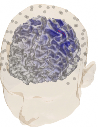
The figure shows the reconstructed activity corresponding SEP responses at Brodmann area 3b found using a hierarchical Bayesian modelling technique, ZI, and a realistic head model. The EEG electrodes attached on the scalp are also illustrated.