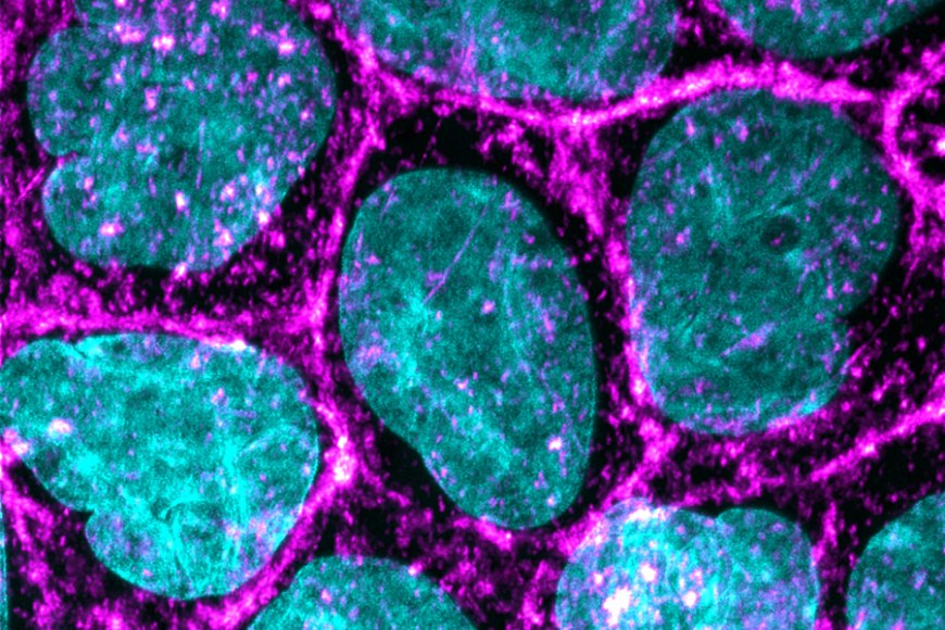
In a collaborative research within ABioT project three research groups demonstrated the prospects of using photoreactive azobenzene-containing DR1-glass to mechanically stimulate cells with fine spatiotemporal control. The nanoscale material deformation upon laser scanning was first thoroughly characterized and then exploited for the mechanical stimulation of epithelial cells. The light stimulation of the DR1-glass caused lateral flow of the material in a laser intensity dependent manner. When combined with cell culture, the light induced deformation of DR1-glass led to strong Ca2+ transients in the epithelial cells which were detected immediately after the stimulation. Real-time monitoring of consequential rapid intracellular Ca2+ signals reveals that the mechanosensitive cation channel Piezo1 has a major role in generating the Ca2+ transients after nanoscale mechanical deformation of the cell culture substrate.
Calcium responses in multiline stimulated sample in normal conditions – advs6609-sup-0004-videos3 (1)
The study was published in November 2023 in the prestigious journal Advanced Science. Read more…

