Written: Miina Björninen
Photo: Jonne Renvall
Creativity challenged for establishing new characterisation methods
Mykuliak & Yrjänäinen tested the ability of mesenchymal stem cells (MSC) to support microvascular network formation, i.e., capillaries, which are the smallest vessels found in nearly every tissue. MSC were isolated from adult human patients from two different locations bone marrow and adipose tissue. The MSC from the different locations were compared for their vessel supporting ability.
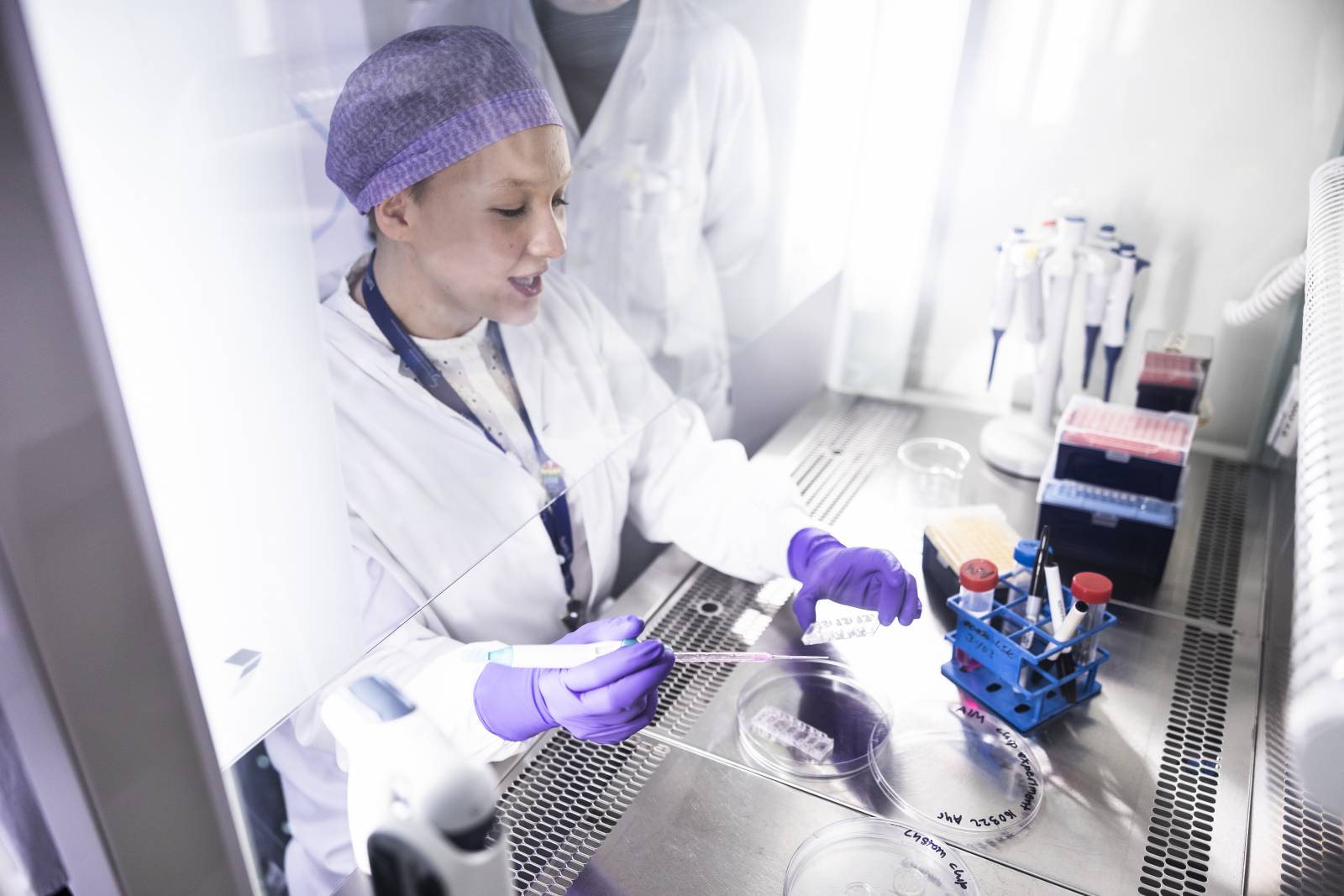
In the laboratory, endothelial cells and MSC were mixed with a fibrin hydrogel and transferred into commercial microfluidic chips. The chip facilitated interstitial flow, across the culture area. Interstitial flow is an important factor in mimicking the flow of blood and tissue fluids in the body.
Results suggested that bone marrow derived MSC supports the formation of larger and more robust vasculature than adipose tissue derived MSC. The field of organ-on-chip is so new, that there is a lack of well-established characterization methods.
“Peer reviewers asked for quantifying interstitial flow but there wasn’t any gold standard method on characterizing the flow in literature. We needed to come up with one” Yrjänäinen recalls.
Together with postdoctoral research fellow, Antti Mäki, they established a whole new characterization method that can be utilized in various experimental set ups.
“Only one functional vessel?”
Mykuliak and Yrjänäinen were thrilled to see for the first time how a test bead was flowing through a newly formed vessel in their 3D vascular model. Months of tedious laboratory work had finally resulted in one functional vessel – out of hundreds or perhaps thousands of not fully formed vessels.
The work was highly multidisciplinary with engineers and cell biologists working together. While success in cell work was often highly unpredictable depending on the nature of the patient derived cells, a stable hand during pipetting and environmental changes, the engineering side was more predictable with a strong emphasis on mathematical modelling.
Therefore, the news of only one functional vessel did not initially impress the engineer colleagues. Luckily, the yield increased upon several optimization rounds. The understanding of each other’s work also grew along the intensive collaboration.
Putting vessels to use
Currently, the ‘vascular duo’ is continuing their own research pathways; Mykuliak is studying how prostate cancer cells escape the blood vessels and enter bone tissue. Yrjänäinen is optimizing a novel chip structure joining together vascular network and mature adipocytes as a 3D adipose-tissue-on-chip model.
Access the full article here.

