


Induced pluripotent stem cells (iPSCs)
Our main focus is to produce patient-specific iPSC lines derived from individuals carrying genetic cardiac disorders such as LQT, CPVT, and HCM. We are reprogramming human somatic cells (skin fibroblasts and blood cells) into iPSCs and characterize those cell lines to produce high-quality iPSC lines for research.
In addition to iPS cell line derivation, we differentiate these patient-specific iPS cell lines into functional cardiomyocytes, cardiac cells, and hepatocytes.
The main techniques:
- PBMC isolation and electroporation for hiPS cell line production
- Human iPS cell line culturing, characterization and hiPSC-CM differentiation
- Immunocytochemistry (pluripotency and hiPSC-CMs)
- PCR/qPCR
- Imaging (video imaging, fluorescent imaging, confocal microscopy)
- Flow cytometry (CytoFlex, FACS Aria)
- Magnetic-activated cell sorting (MACS)
- Seahorse metabolic analysis
- Ca2+-imaging
Cardiac ischemia modeling
Our group has extensive experience in cardiac ischemia modeling using organ-on-chip platforms to study the mechanisms of ischemia–reperfusion injury and to develop novel therapeutic strategies and treatment options. Cardiac ischemia remains a major contributor to global mortality, highlighting the need for research aimed at improving our understanding of its underlying mechanisms and pathophysiology. Using our current ischemia modeling platforms, we can generate either uniform hypoxic conditions for hiPSC-CM monolayers and 3D cardiac tissues, such as engineered heart tissues (EHTs), or controlled oxygen gradients across hiPSC-CM monolayers. These platforms enable the investigation of multiple aspects of cardiac ischemia and provide versatile tools for studying disease mechanisms and evaluating potential therapeutic interventions.
Main techniques
- Ischemia-on-chip and Ischemia Border Zone-on-chip
- Physioxic oxygen environment
- Unifor hypoxia and reoxygenation
- Oxygen gradient and reoxygenation
- Oxygen monitoring: live cell hypoxia dye & ratiometric luminescence-based oxygen imaging
- Microelectrode array technology
- Video-based functional analysis
- Immunocytochemical staining
- RT-qPCR
- Calcium imaging
Cardiac organoids for modeling cardiomyopathies
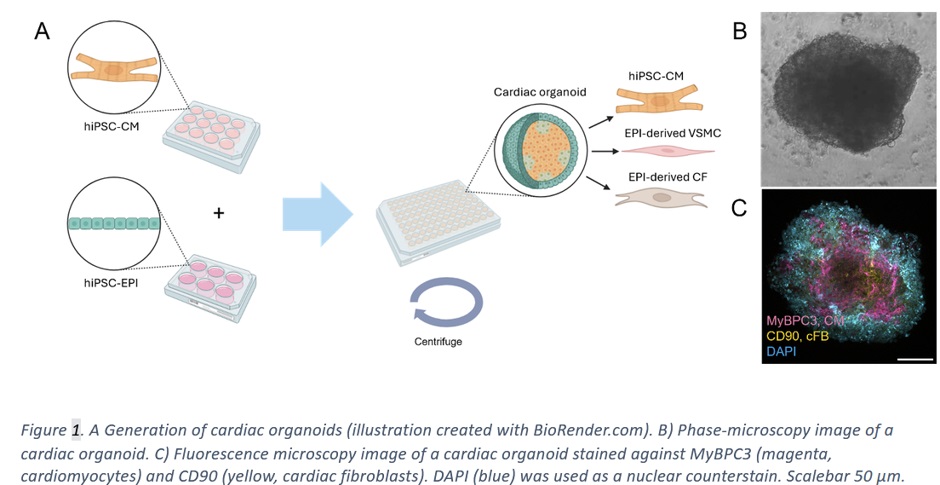
Cardiac organoids provide a medium-throughput platform of more physiologically relevant tissue constructs for modeling both genetic and acquired cardiomyopathies. They allow more accurate recapitulation of disease mechanisms, especially in the context of crosstalk between different cell types of native cardiac tissue, including cardiomyocytes, cardiac fibroblasts and vascular smooth muscle cells. Currently, Heart group utilizes the organoids to model hypertrophic cardiomyopathy caused by a Finnish founder mutation in junctophilin-2 gene.
Main techniques
- hiPSC maintenance
- hiPSC differentiation into cardiomyocytes and epicardial cells
- Magnetic-activated cell sorting (MACS)
- Flow cytometry
- Immunocytochemical staining
- qRT-PCR
- Western blot
- Video-based functional analysis
- Calcium imaging
Engineered heart tissue
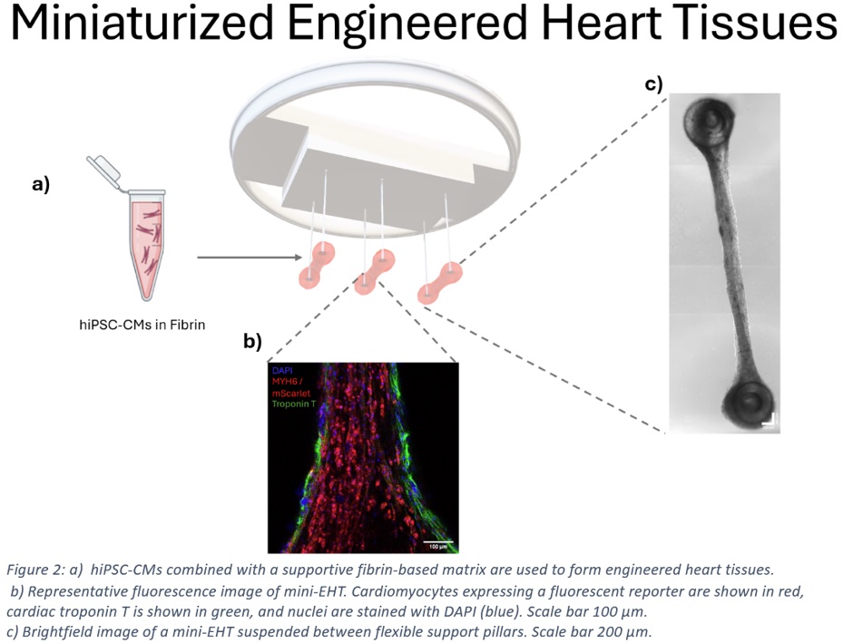
Engineered heart tissues (EHTs) are three-dimensional cardiac models created by combining human induced pluripotent stem cell-derived cardiomyocytes (hiPSC-CMs) with a supportive biomaterial matrix. Within the tissue, the cells self-organize into a synchronized, contractile structure that more closely resembles native human myocardium than conventional two-dimensional cell cultures.
In our research, we develop and utilize miniaturized EHT models that enable efficient generation of human cardiac tissues while reducing cell consumption and increasing experimental throughput. By combining advanced imaging, automated data analysis, and functional measurements, we characterize cardiac tissue development, maturation, and function. The platform is also applied to disease modeling, including studies of hypertrophic cardiomyopathy (HCM), providing insights into disease mechanisms and potential treatment strategies.
Main techniques
- hiPSC maintenance
- hiPSC differentiation into cardiomyocytes
- Magnetic-activated cell sorting (MACS)
- Immunocytochemical staining
- Video-based functional analysis, Contractile force measurements
- Calcium imaging
- Confocal microscopy
Cardiac innervation model
Neuro-Cardiac Organ-on-Chip
The neuro-cardiac organ-on-chip is a human stem cell-based in vitro model designed to investigate interaction between the nervous system and the heart under both physiological and disease conditions. By combining human induced pluripotent stem cell (hiPSC)-derived cortical neurons, sympathetic neurons, and cardiomyocytes within a microfluidic platform, the model recreates key aspects of the brain-heart axis in a controlled environment.
This platform enables the study of how neuronal activity influences cardiac function and how disruptions in neurocardiac communication contribute to cardiovascular diseases such as CPVT. In addition to disease modelling, the system provides a powerful tool for investigating cellular mechanisms, identifying therapeutic targets, and evaluating drug responses in a human-relevant setting.
Key techniques
- Human iPSC culture
- Microfluidic organ-on-chip technology
- Compartmentalized multiculture of neurons and cardiomyocytes
- CPVT-patient derived cell types
- Multi-level validation of the platform
Liver model
Developing an in vitro liver model for cardiovascular disease research
We are developing a liver model that could help to unwind the liver-related pathways involved in the increased risk of plaque rupture and acute CAD phenotype.
Liver has a central role in lipid metabolism and is the major site for generation of plasma lipids and lipoproteins. Thus, a mature in vitro liver model recapitulating the native liver architecture and functionality is useful in studying the effect of different lipids and inflammatory proteins on atherosclerosis, but it could also be utilized in drug screening and hepatotoxicity studies.
We utilize induced pluripotent stem cells (iPSCs) from patients with different coronary artery disease (CAD) phenotype (stable disease / acute disease / healthy) and differentiate them to hepatocytes. We have used the iPSC-derived hepatocytes in 2D for projects that model inherited traits of lipid metabolism in CAD patients. Furthermore, we have extended our studies into miRNA and proteomic profiling of the iPSC-hepatocytes in different patient groups.
Currently, we are working towards a 3D liver in vitro model that we can utilize for different aspects of cardiovascular disease research. We use iPSC-derived hepatocytes and culture the cells either on commercially available or custom-made microfluidic chips. We utilize various biomaterials in these cultures to support the formation of a 3D microenvironment and promote liver functionality.
The main techniques
- Cell culturing and iPSC to hepatocyte differentiation
- Flow cytometry (CXCR4 for DE cell indentification)
- Culturing cells on microfluidic chips
- Immunocytochemistry
- ELISA assays for measuring e.g. albumin and urea production
- QPCR (mRNA and miRNA)
- Imaging (EVOS FL, Confocal, SPIM)