Our team conducts research at the interface of Chemistry, Material Science, and Biological Sciences on the following topics.
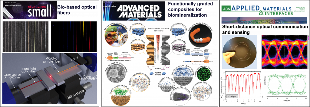
Bio-based materials for optics and photonics
Increased use of plastic-based materials in consumer products will amplify the number of microplastics that eventually end up in the food chain. We utilize naturally abundant, renewable, and sustainable molecular, polymeric, and colloidal-level building blocks. We aim to provide alternative and innovative solutions to overcome fossil fuel-based materials. Through this, we provide a transition to green and sustainable materials and processing technologies for bio-based optics, photonics, and health technology. Our research objectives support the climate-neutral policies, circular economy, European Union Green Deal, and United Nations sustainable development goals by effectively using material stock and flow and creating a new value chain. We use biopolymers for optomechanically tunable gels, optical fibers, and fiber lasers.

Related publications: Small 2021, Adv. Mater. 2021, Adv. Mater. 2021
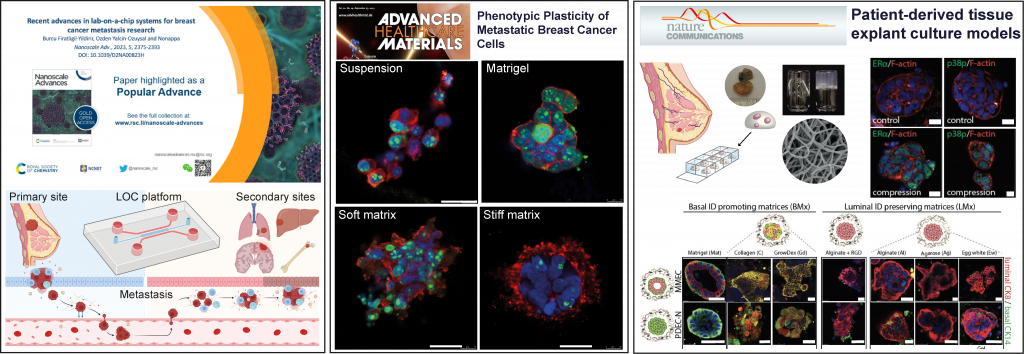
Breast cancer disease and diagnostic models
As of 2020, breast cancer is the most frequently diagnosed cancer type and is one of the leading causes of cancer-related deaths in women. Despite the high incidence of the disease, the average 5-year survival rate of breast cancer patients with early-stage, non-metastatic disease is over 80%. However, this survival rate falls below 25% in patients with metastasis. Metastatic breast cancer is therefore considered practically incurable and is responsible for more than 90% of tumor-related deaths, often due to impaired vital organ functions. Our team is developing lab-on-a-chip (LOC) system-based in vitro models to study the multistep and complex metastasis process. The ultimate aim is to develop a suitable model to study breast cancer metastasis cascade and a preclinical model for metastatic cancer treatment. We actively develop hydrogel-based three-dimensional extracellular matrices (ECM) mimic for a cancer cell, tissue, and explant culture. We utilize molecular and polymer hydrogels with unique rheological properties, chemical functionalities, and biochemical cues to study cell-matrix interactions. Particularly, we mimic the non-linear viscoelasticity of biological systems in artificial scaffolds. The hydrogels are used for 3D tissue culture, patient-derived tissue cultures, and organoid culture.

Related publications: Nanoscale Adv.2023; Adv. Healthcare Mater. 2023; Nat. Commun. 2021,
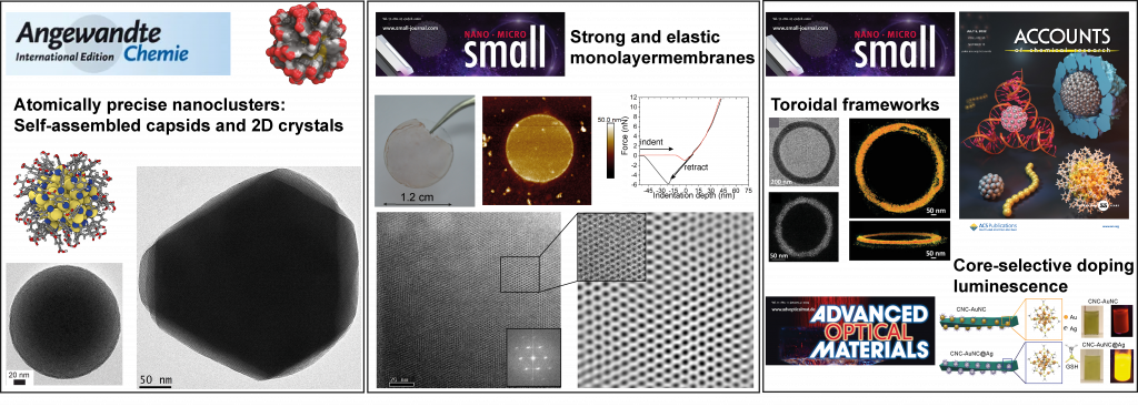
Precision nanomaterials
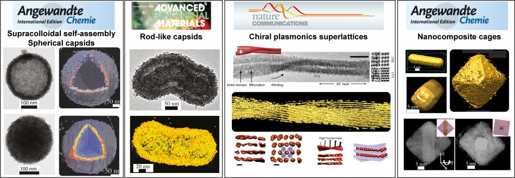
We use atomically precise noble metal (gold and silver) nanoclusters and narrow-size dispersed plasmonic metal nanoparticles. We are developing concepts and methods for precision nanoparticle self-assemblies across length scales. We tune the functionalities of the organic ligands to control the inter-nanoparticle interactions. Specifically, we use hydrogen bonding, electrostatic interactions, and metal coordination as major driving forces or combinations of more than one interaction. We are developing one-dimensional nanowires, 2D colloidal crystals, or 3D colloidal frameworks. We amplify optoelectronic properties and mechanical performance through self-assembly and develop approaches for various composite chiral plasmonic superstructures.

Related publications: Small 2023, Small 2022, Small 2021, Angew. Chem. Int. Ed. 2018, Angew. Chem. Int. Ed. 2016
Advanced transmission electron microscopy (TEM)
We use advanced TEM imaging, sample preparation, and image processing methods. Specifically, we focus on cryogenic transmission electron microscopy (Cryo-TEM), electron tomography (ET), in situ solid state, and liquid cell TEM.
Cryogenic-TEM: Transmission Electron microscopy (EM) is a powerful imaging technique to study a diverse range of synthetic and biological materials. Biological samples or synthetic soft nanostructures lead to numerous drying artifacts and collapsed structures. The cryogenic sample preparation method allows imaging of the biological materials and soft nanomaterials near their native or frozen states with reduced artifacts. The specimens are rapidly frozen using a liquid propane/ethane mixture, resulting in thin (below 130 nm) and transparent vitreous ice. This method suits a broader range of particles, such as virus particles, polymer micelles, vesicles, nanotubes, and protein assemblies.
Electron Tomography: TEM images of three-dimensional (3D) objects produce superimposed two-dimensional (2D) orthogonal projections. Importantly, in transmission electron microscopy (TEM) images, the 2D projection often contains high-resolution structural details. A limited amount of correct internal structural information is obtained from the 2D images because of the superimposed nature. We use electron tomography to achieve 3D reconstruction using a series of 2D projections to overcome the above limitations. Electron tomography thus allows the most realistic and high-resolution structural and morphological features of nanostructures near-atomic resolution.

Related Publications: Angew. Chem. Int. Ed. 2020 ACS Nano 2019 Angew. Chem. Int. Ed. 2018, ACS Nano 2019 Adv. Mater. 2019 J. Am. Chem. Soc. 2018 Nat. Commun. 2017
