
X-ray Microtomography
Jari Hyttinen ![]()
Markus Hannula
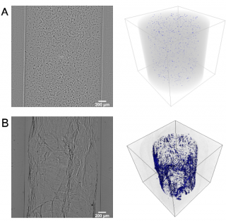
X-ray microtomography (µCT) is a means to examine the internal 3D structure of an object in micrometer-scale. The method is based on X-ray attenuation, hence it is can also be applied to opaque objects. The contrast arises from the density variation of the structures and can be enhanced with contrast agents.
We are developing µCT methods especially for tissue engineering applications. It is a valuable method in development and quality control of (bio)materials and we are working hard to develop tools, not only for material research but also for tissue engineering research. µCT is a widely used method in bone research due to the excellent contrast of calcified tissues. Soft tissues, however, are challenging because the density of different soft tissues is relatively close to each other and that of water, which leads to poor X-ray contrast. In order to enhance the soft tissue contrast, we are developing methods to transport heavier elements into tissues both as background stains and specific antibody-based stains for even cellular imaging using X-ray microtomography. Moreover, our microtomography equipment supports in-line phase contrast imaging, which can also be used for contrast enhancement.
3D Optical Imaging
Biophotonics science deals with interaction between photons and biological matter with a range of applications that extends from medical diagnostics to therapy and disease prevention. We are interested in biophotonics technologies applied for imaging in the mesoscopic regime, i.e., biological samples with sizes between 1 mm and 1 cm – especially those of tissue engineered grafts.
We are building a hybrid imaging tool based on optical projection tomography (OPT) and selective plane illumination microscopy (SPIM) for image 3D tissue engineered constructs over time. Our research aims is to develop real-time, non-invasive and non-destructive methods for live and high-resolution imaging of 3D TE products over time. Imaging techniques for characterization prior to application 3D structure of scaffolds, as X-ray µCT, is relatively well-developed, and it is being used for TE imaging. The hybrid imaging tool being developed will able to assess the morphology, location, differentiation and function of the cells within the 3D-biomaterials, helping in the evaluation of engineered tissue for regenerative therapies and for tissue models.
Texture Analysis
Michelangelo Paci ![]()
Antti Ahola
We develop texture descriptors and ensembles of classifiers as tools for cell and tissue classification and assessment. Extracting textural information from images has be proven to be a successful approach for many image processing and pattern recognition tasks, such as segmentation and classification exploiting very diverse kind of images, from single cell to CT/MRI images. Our research aims to use of texture analysis to assess different features of biological tissues, such as their quality or their maturation level, in order to provide our labs appropriate software tools.
Selected Publications
- Nanni, Paci, et al. An ensemble of visual features for Gaussians of local descriptors and non-binary coding for texture descriptors. Expert Syst Appl. 2017;82:27-39. [ScienceDirect]
- Nanni, et al. Ensembles of dense and dense sampling descriptors for the HEp-2 cells classification problem. Pattern Recognit Lett. 2016;82(P1):28-35. [ACM]
- Nanni, Paci, et al. Texture Descriptors Ensembles Enable Image-Based Classification of Maturation of Human Stem Cell-Derived Retinal Pigmented Epithelium. PLoS One. 2016;11(2). [PubMed]
- Nanni, Paci, et al. Analysis of virus textures in transmission electron microscopy images. Stud Health Technol Inform. 2014;207:83-91. [PubMed]